Synchrotron
X-ray studies at Argonne National Laboratory
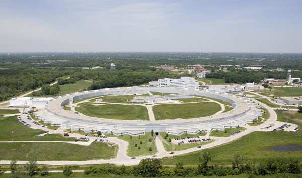 Areal View of the Advanced Photon Source at Argonne National Laboratory Areal View of the Advanced Photon Source at Argonne National Laboratory
Several advanced X-ray
techniques are utilized at the Advanced Photon Source in Argonne
National Laboratory for measuring crystal quality, structure, and
magnetic properties of our thin-film samples. Due to the small amount
of material deposited through MBE and the spatially dependent
composition of these combinatorial samples, table-top x-ray sources are
largely underpowered to accomplish the needed high-intensity
micron-sized beam. In addition, synchrotron sources are ‘white’
sources, allowing energy-dependent measurements to be made – something
impossible from table-top x-ray sources. In the figure below, both the
intensity and energy-dependence advantages of the source is obvious.
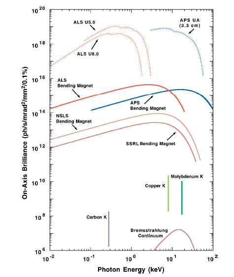 Source
brilliance versus energy for various facilities in the world including
the Cu Ka lines' brilliance, standard in table-top sources. Source
brilliance versus energy for various facilities in the world including
the Cu Ka lines' brilliance, standard in table-top sources.
Combinatorial X-ray Diffraction (XRD)
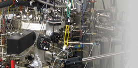
All
structural studies at the APS are conducted in collaboration with Dr.
Yong S. Chu. His beamline contains a double-crystal monochromator with
an energy band-pass of 0.01%, Kirkpatrick-Baez mirrors capable of
wavelength-independent x-ray focusing to 3 microns, a Huber 4-circle
diffractometer with angular precision to 1x10⁻⁷ radians, a Si
drift-diode detector/multi-channel analyzer for taking spectra, and a
variety of scintillation detectors and cameras (see diagram below). All
of this equipment is ideal for the combinatorial samples produced at in
our lab, allowing high throughput characterization of crystal structure
and quality as a function of composition. For example, at each point on
a sample, crystallographic information can be taken using standard
x-ray diffraction techniques via a NaI scintillator and diffractometer,
while the composition can be simultaneously monitored using x-ray
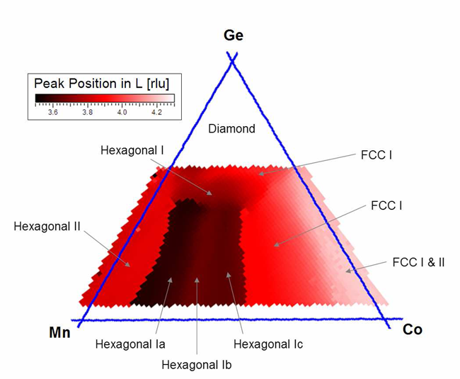
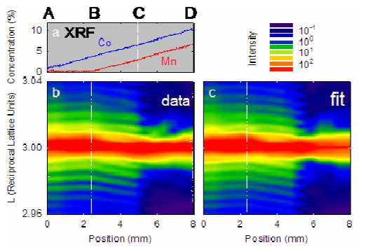
florescence via the Si drift-diode detector. In the figure, the
crystallographic phase of a CoxMnyGez ternary sample was determined at
all compositions.
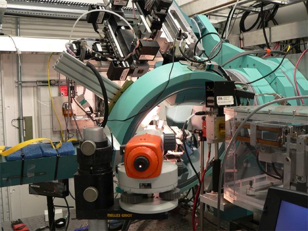
Crystal Truncation Rod Analysis
Among
the advanced techniques used at the APS is Crystal Truncation Rod (CTR)
Analysis. Due to the high quality of epitaxial films, the atoms in the
film makeup 2D planes (like a horizontal grating) on the surface of the
substrate. These atomic planes have a different lattice spacing than
the substrate due to dopants we introduce during growth of the film or
because the film is of different elemental species than the substrate.
This can be interpreted as strain if the film atoms are coherent with
the substrate atoms in-plan and can be detected as a fringe pattern
when this film grating diffracts with the grating made up by the
substrate atoms. From this mechanism, the strain states and/or the
lattice parameter can be measured very precisely. Furthermore,
Debye-Waller disorder and film thickness can be obtained from other
parameters of the fringes. As composition varies, these physical
parameters will vary and their trends can shed light into the
mechanisms controlling material properties on the macroscopic level.
 CTR
data and fits, which extract strain information from the position of
the interference fringes with respect to the main Bragg reflection at
the center. CTR
data and fits, which extract strain information from the position of
the interference fringes with respect to the main Bragg reflection at
the center.Energy-Dependant
TechniquesA second group of techniques involves
inelastic scattering and allows for more localized information
regarding structure and disorders. X-ray Anomalous Fine Structure
(XAFS) is a spectroscopic technique that probes an atom’s neighboring
atom elemental type, coordination number and bond distance. The
technique involves systematically changing the incident beam’s energy
across an atom’s spectral excitation (unique to each element) and
monitoring its absorption of x-rays. The fine oscillations in
absorption as a function of energy (frequency) can be Fourier
transformed into real-space to give peaks representing neighboring
atoms at specific distances. A more difficult but more rewarding
technique utilized at the APS is Diffraction Anomalous Fine Structure
(DAFS). This time, the absorbed intensity of a Bragg reflection is
monitored, which gives site-specific local information rather than just
element-specific. More generally, anomalous diffraction (DAFS minus the
fine oscillations) can illuminate site occupancy and crystallographic
site swapping between elements. Put together, the chemical ordering and
specific types of chemical disordering within a crystal can be measured
precisely. 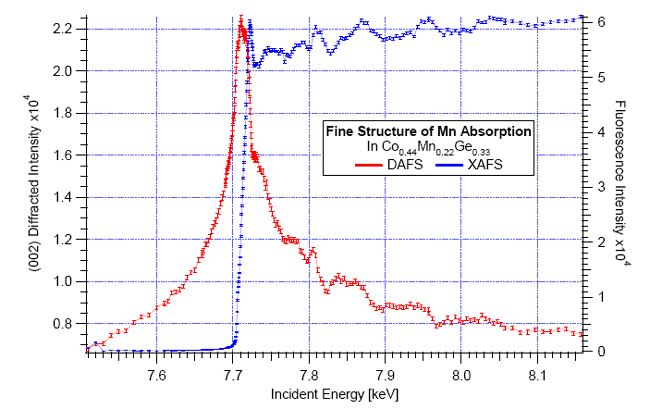 An
example of DAFS and XAFS data which can be fit to reveal the local
structure around specific atoms in the matrix, in this case the Co
atoms. An
example of DAFS and XAFS data which can be fit to reveal the local
structure around specific atoms in the matrix, in this case the Co
atoms. |