Home | People
| Projects | Techniques
| Publications | Contact
| Links
| Advanced
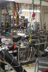
Combinatorial Molecular
Beam Epitaxy (MBE) MBE
is a set of versatile techniques for synthesizing and characterizing
epitaxial structures with precise control. The low energy molecular or
atomic beams are produced thermally in ultrahigh vacuum (UHV), and as
the molecules or atoms land on a heated substrate surface facing the
beams, nanostructures form. What distinguishes MBE from other deposition
techniques are its precise control of deposition parameters and the
available in-situ characterization techniques. Combinatorial MBE is a
powerful approach we practice that synthesizes and characterizes a
large number samples with a range of compositions and growth parameters
on a single substrate.
Read
more...
| | | 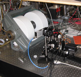 | Magnetooptic Kerr
Effect (MOKE) The Magnetooptic Kerr Effect is used to study
the magnetic
properties of our samples. When linearly polarized laser
light
reflects off a magnetized sample, it undergoes a change in its
polarization state that can be measured. This change is
caused by
off-diagonal elements in the dielectric tensor of the material as a
consequence of spin-orbit coupling.
Read
more... |
| | 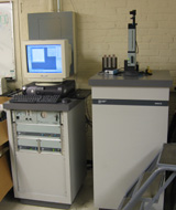 | Superconducting Quantum Interference Device (SQUID)
The SQUID magnetometer at UNC is a
Quantum Design MPMS system. It can
measure from 2-400K in fields upto 5.5 Tesla. A large variety of
samples
are investigated using this system, including GMR and CMR materials,
Carbon nanotubes, Heusler alloys, metallic spin-glasses and others. |
| | 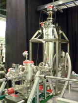
| UHV
Magnet Cryostat
Within this
Janis-built UHV
magnet cryostat, scanning probe microscopy (SPM)/spectroscopy and
magnetooptical
measurements are conducted in a temperature range between 1.6K and 350K
and a
field range of up to 3 Tesla. The instrument is capable of simultaneous
microscopy and spectroscopy techniques in a variety of imaging and
metrology
modes including tunneling, topographic, dielectric
constant/conductance, and
magnetic resonance, using scanning tunneling microscopy and evanescent
microwave probe microscopy. The
precision xyz sample stage with a large travel range (1.5 cm) allows
SPM
measurements to be conducted over an entire ternary combinatorial
sample. The
instrument makes it possible to probe
nanoscale surface structure, magnetism, and electronic states,
including
quantitative measurement of spin polarization using superconducting
point contact Andreev
reflection.
| | | 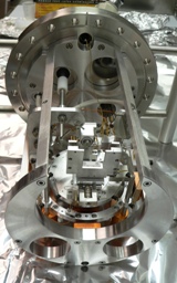 | Scanning
Tunneling
Microscopy (STM)
This
is a room temperature UHV scanning probe microscope (SPM) (Park
Scientific SPM
VP). It is capable of both STM and AFM for in-situ studies of surface
structure
and morphology of MBE-grown materials. |
| | 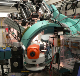 | Synchrotron
X-Ray Studies
at Argonne National Lab (XRD) Synchrotron
radiation is used to carry out x-ray diffraction/spectroscopy
experiments at
the Advanced Photon Source in Argonne National Laboratory. Its high intensity,
micron-sized beam allows
for studies of structural properties and composition of combinatorial
samples,
using x-ray diffraction and fluorescence spectroscopy techniques. The source’s tunable
energy is also used to
probe local structures, such as chemical order, oxidation state, and
bond
length.
Read
more... |
| | 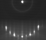 | Reflection High Energy
Electron Diffraction (RHEED)
Reflection High Energy
Electron Diffraction (RHEED) has been
the work horse for real-time quantitative
characterization of epitaxial
growth, owing to its surface sensitivity, large working distance, and
the
reflection geometry compatible with deposition. The grazing incident
angle for
the RHEED beam with respect to the surface, typically 1-2 degrees gives
rise to
a very small penetration depth, resulting in a diffraction pattern that
is
sensitive to only the outer most surface layer. Therefore, by
monitoring the RHEED
patterns (diffracted electrons) during growth and across the sample,
evolution
of surface structures can be examined systematically.
"Rheed"
more...
|
|